


The COVID-19 pandemic has undoubtedly changed the course of countless lives. And for many of those who have contracted the disease, the effects can still be felt even if they’ve “officially” recovered.1
Commonly known as long COVID, this side effect of the SARS-CoV-2 infection affects millions worldwide. In a study of sailors on a French aircraft carrier, more than half of those who’d had COVID still reported symptoms a year later, although 40% of sailors who had tested negative reported similar symptoms at that point.2 For a few years, researchers have been hitting dead ends as to what causes these long-term issues. One study has added evidence supporting one of their leading theories — microclots.3
Research Deepens Understanding of Long COVID
In a study published in the Journal of Medical Virology, researchers set out to learn what makes long COVID so persistent. Building on their earlier work, they focused on the blood itself and found that those with long COVID carry more and larger abnormal microscopic structures. Specifically, fibrin microclots linked to immune cell debris.4
To perform the analysis, the team used advanced imaging to examine how the microclot structures are built, what they contain, and how they differ from healthy blood. For the study population, they selected 50 adults with long COVID who continued to struggle with problems such as exhaustion, shortness of breath, brain fog, dizziness, and chest tightness long after the initial infection had been resolved. They compared these patients with 38 healthy controls.
• There were stark differences in blood structure — According to the researchers, the test samples had far more microclots than healthy controls (a nearly 20-fold difference in the median count), and the clots tended to be larger.
Digging deeper into the data, they discovered that the microclots were intertwined with neutrophil extracellular traps, or NETs. These are sticky webs of DNA and enzymes that neutrophils release when activated. The researchers suggest that NET-rich clots may resist normal breakdown processes, which could leave lingering blockages at the microvascular level.
• The impact of NETs — The researchers reported major increases in several NETs markers — myeloperoxidase, neutrophil elastase, and circulating cell-free DNA — in the long COVID group. These are enzymes and DNA fragments that may act like glue inside the clots. The more NET material the researchers found, the more microclots there were.
• Another noteworthy detail is the size of the structures — Imaging showed that 60% of long COVID patients had the largest microclots (over 1,600 µm²), compared with 13% of healthy controls. Healthy controls also had microclots, but they were fewer and smaller.
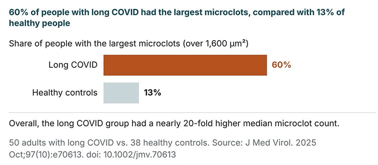
Figure 1. People with long COVID had a nearly 20-fold higher median microclot count, and 60% had the largest microclots, compared with 13% of healthy controls.
• How deeply embedded the NET components were within the clots — Microscopy showed DNA and the neutrophil enzyme myeloperoxidase within the microclots, not just around them, although the authors noted this method can’t reliably measure what proportion of microclots contain them. This is important because fibrin-based clots normally break down with the help of enzymes like plasmin.
The researchers suggest that when DNA and neutrophil proteins are built into a clot, the breakdown process may become inefficient and slow, which could help explain why long COVID can last for months or even years.
• Microclot size mattered — For example, all three NET markers rose along with microclot counts, and microclots in the 100 to 400 µm² range, the most common size, tracked most closely with them. The authors suggest these markers could one day help diagnose long COVID. It may also help researchers sort patients into subgroups in the future.
• NETs act as scaffolding — When neutrophils eject their DNA and enzymes, they create long, sticky strands designed to trap pathogens. In long COVID, the researchers propose that these strands latch onto fibrin — a protein involved in clotting — and build layered, tangled structures. Once formed, they may float through the bloodstream, lodging in tiny vessels and creating micro-obstructions. The result could be impaired microcirculation and reduced oxygen transfer.
• Another mechanism the researchers proposed involves the resistance of these clots to fibrinolysis — Because NET components may stiffen and reinforce the fibrin structure, the body’s usual cleanup systems may struggle to dissolve them. This could lead to chronic retention of microclots, continued vascular stress, and prolonged symptoms.
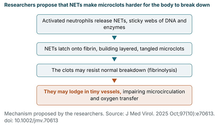
Figure 2. The researchers propose that NETs bind to fibrin, forming microclots that may resist breakdown and lodge in tiny blood vessels.
• The amount of NET material increased directly alongside the amount of microclots — This suggests the immune and clotting systems may be interacting in a loop, each one pushing the other. Over time, such a loop could drain your energy, stress your cardiovascular system, and keep inflammation alive long after the virus is gone.
As the authors note, an association like this doesn’t prove cause and effect. They also didn’t compare long COVID patients with people who have other diseases, such as diabetes, sepsis, or autoimmune disorders, so it’s not yet clear how specific these changes are to long COVID.
You’re Already at Risk of Blood Clots Once You Get Infected
In an earlier study published in Hematology Reports, researchers reviewed evidence on how the SARS-CoV-2 virus can trigger dangerous clotting problems during the infection and long after someone tests negative. Their goal was to identify how the virus flips the body into a clot-forming state, why this risk lasts for months, and which biological events drive the damage.5
The researchers reviewed how both acute and long-term clotting problems interfere with blood flow, stress the cardiovascular system, and set the stage for health issues that may overlap with symptoms seen in long COVID.
• Rates of clotting complications — Deep-vein thrombosis and pulmonary embolism increased dramatically among hospitalized COVID patients, and the researchers reported that in one study, nearly a third of critically ill patients still had blood that clotted too easily even while receiving anticoagulant therapy designed to prevent clots. Even so, another study identified preventive anticoagulant therapy as a “protective factor” against pulmonary embolism.
• What happened after people left the hospital — Extended clotting risk showed up in multiple studies, with patients showing clot formation weeks to months after the initial infection had passed.
• A comparison of clot complications across different groups — The authors noted that those with preexisting cardiovascular disease, diabetes, obesity, or advanced age were more likely to experience severe clotting events. Clotting complications during the acute illness were uncommon in people with mild illness who weren’t hospitalized. However, one large study cited in the review found that clot risk was still raised nearly a year later, including in people who were never hospitalized.
• SARS-CoV-2 creates imbalance between clot formation and clot breakdown — Under normal conditions, your body constantly forms tiny clots and dissolves them. But according to the researchers, COVID disrupts this balance by releasing clot-promoting chemicals, damaging vessel walls, and activating platelets at unusually high rates. Once this imbalance begins, your body forms clots faster than it dissolves them.
• Biological mechanisms that tie the processes together — The paper explains how the virus may infect endothelial cells, which are the thin linings inside your blood vessels. When these cells are damaged, they release substances that encourage clotting, narrow the vessel opening, and attract platelets to the injury site.
The result is a chain reaction — damaged endothelium signals inflammation, inflammation triggers platelet activation, and activated platelets send more clotting signals.
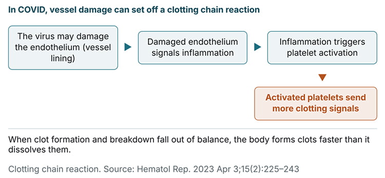
Figure 3. In COVID, damage to the vessel lining can set off a chain reaction of inflammation, platelet activation, and more clotting signals.
• The immune system’s involvement — COVID-19 stimulates a wave of inflammatory cytokines, which are chemical messengers that tell the body to mount a defense. When these cytokines rise too high, your clotting system receives constant signals to activate.
Some researchers think this may be why some people feel as if their bodies never shut off even after the infection is gone — their inflammatory and clotting pathways may remain switched on, draining energy and straining the heart, lungs, and muscles.
• How platelet behavior changes during and after infection — Platelets are tiny cell fragments that help your blood clot when you’re injured. According to the researchers, the SARS-CoV-2 virus drives platelets into a highly activated state where they clump together more easily, interact abnormally with immune cells, and release clot-promoting substances.
Researchers have proposed that this heightened reactivity may not return to normal right away, and that ongoing platelet activation could contribute to lingering symptoms, even when scans or standard tests look fine.
Strategies to Help Manage Long COVID Better
Mitochondrial dysfunction is thought to play a key role in long COVID. If these energy factories inside your cells don’t function at an optimal rate, your energy levels will ultimately be affected. If you know someone who is suffering from this condition, these strategies may help support recovery. Talk with a healthcare provider before starting any of them, especially if you take medications:
1. Proteolytic enzymes — Whenever I encounter someone who is experiencing long COVID symptoms, one of the first things I suggest is proteolytic enzymes, such as nattokinase, lumbrokinase, and serrapeptase, which may help clear residual spike protein from the body, ease inflammation, and give the brain and tissues space to recover. In a laboratory study, nattokinase broke down the SARS-CoV-2 spike protein in cell cultures,6 but this hasn’t been tested in people with long COVID.*
These enzymes are best taken on an empty stomach, at least an hour before, or two hours after, consuming protein. If taken with food, they may be diverted into digesting your meal instead of clearing problematic proteins from the bloodstream. Because these enzymes break down fibrin, they may increase bleeding risk. So if you take a blood thinner or antiplatelet medication, have a bleeding disorder, or have surgery scheduled, talk to your doctor before using them.
*These findings are from laboratory or animal research and may not directly apply to human health.
2. Consider the I-RECOVER protocol — The Independent Medical Alliance (IMA), previously known as the FLCCC, offers a detailed plan called the I-RECOVER protocol. It’s a detailed guide for addressing post-vaccine and post-infection complications.
It outlines strategies for detoxification, calming inflammation, and restoring mitochondrial function — all foundational steps for improving health after COVID-related injuries. Several of its recommendations involve prescription medications, so work through it with a healthcare provider.
3. Reduce intake of linoleic acid (LA) — Your mitochondria rely on a unique lipid called cardiolipin,7 which is located within the inner membrane structures (cristae) where energy is produced. It stabilizes the electron transport chain, maintains the folded cristae structure that expands surface area for energy production, and helps manage proton flow needed to generate ATP.
This is more important than most people realize, as the wrong dietary fats can distort cristae structure and hinder energy production. Roughly 20% of this membrane is composed of cardiolipin, a distinctive phospholipid that contains four fatty acid chains instead of the usual two.
Importantly, cardiolipin requires LA to function properly. The problem begins when excess dietary LA accumulates in the other 80% of the inner mitochondrial membrane — phospholipids that are not designed to carry high LA loads. In those surrounding membranes, LA is highly prone to oxidation.
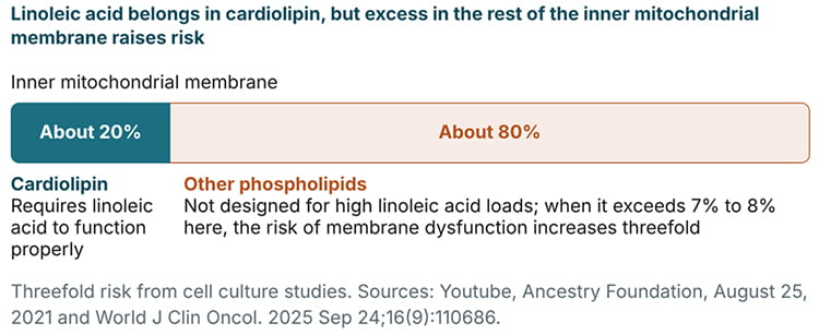
Figure 4. Cardiolipin makes up about 20% of the inner mitochondrial membrane and requires linoleic acid; excess linoleic acid in the other 80% raises the risk of membrane dysfunction.
Reactive oxygen species (ROS) generated during normal energy production initiate lipid peroxidation, forming toxic aldehydes that bind to respiratory proteins, impair electron transfer, and amplify oxidative stress in a self-perpetuating cycle. When LA in these non-cardiolipin phospholipids exceeds 7% to 8%, the risk of membrane dysfunction increases threefold.8
Keeping daily LA intake under 5 grams, and closer to 2 grams when possible, helps limit excess incorporation into these non-cardiolipin membrane compartments. LA is found in excess in seed oils (which are used heavily in ultraprocessed and restaurant foods), condiments, nuts and seeds, adulterated olive and avocado oils, and animal products from grain-fed livestock such as conventional chicken and pork. To track your intake, I recommend downloading my PAX app once it’s available.
4. Methylene blue — Some practitioners use this compound for the exhaustion and neurological challenges that linger after COVID infection or getting the shot, although it hasn’t been studied for long COVID. It functions as an electron carrier, essentially acting like a rechargeable energy source. Unlike many compounds that perform a similar role, it’s less likely to promote harmful oxidative reactions at low doses.
When mitochondrial respiration or oxygen utilization is impaired, methylene blue can reroute electron flow, helping sustain energy production even when normal pathways are blocked. It may also help compensate for reduced blood flow by improving mitochondrial efficiency when tissues aren’t receiving enough oxygenated hemoglobin.
Its benefits may extend beyond the time it remains in the body — methylene blue may induce biochemical changes that enhance oxygen-processing pathways and may even promote the formation of new mitochondria over time.
Dosing is hormetic. Low amounts provide benefits that high amounts can negate. Most experts recommend relatively high doses for longer-term treatments, such as 0.5 milligrams (mg) to 1 mg per kilogram of body weight for cognitive support. For a person weighing 150 pounds, that would be a dose of 34 to 68 milligrams. I believe this is excessive and unnecessary.
Doses of more than 3 to 5 milligrams are likely never needed unless you are undergoing treatment for a life-threatening condition such as carbon monoxide or cyanide poisoning. The average dose for most adults that reduces reductive stress is only 5 mg, once a day, regardless of weight. It has a half-life of over twelve hours and will gradually build up if you take it every day, so higher doses are not needed.
Only use pharmaceutical-grade methylene blue in capsule or tablet form, prescribed by a healthcare professional and filled through a compounding pharmacy, as industrial and chemical varieties frequently contain contaminants like heavy metals. Taking it with vitamin C may enhance absorption.
Keep in mind, though, that methylene blue can cause a dangerous reaction called serotonin syndrome when combined with SSRIs and other serotonergic medications, and it isn’t safe for people with G6PD deficiency.
To learn more, read “The Surprising Health Benefits of Methylene Blue.” It features an interview with methylene blue expert Francisco Gonzalez-Lima, Ph.D., who has spent many years studying this drug.
5. Check your iron levels — Excess iron is another major disruptor of mitochondrial function, and many people — aside from menstruating women or individuals with significant blood loss — tend to accumulate too much. In contrast, copper deficiency may be more common than many people realize. A ferritin blood test shows where you stand; the ideal range is 60 to 75 ng/mL.
Iron and copper need to be in balance because they depend on each other for proper metabolism. Copper is required to move and recycle iron efficiently, so low copper can contribute to iron problems such as anemia, even when iron intake is adequate. Copper is also essential for mitochondrial energy production, detoxification, and overall metabolic health.9 You can learn more about this in “The Poorly-Understood Role of Copper in Anemia.”
You can increase copper by supplementing with 4 to 10 mg of copper bisglycinate daily or by consuming copper-rich foods like bee pollen, grass fed beef liver, and acerola cherry (which contains the copper-dense enzyme tyrosinase). The upper limit for adults is 10 mg a day from food and supplements combined,10 so keep your total at or below that and check with your healthcare provider before supplementing.
To reduce excess iron, routine blood donation is a simple and effective method. This usually involves donating two to four times annually, or having smaller amounts removed monthly by a healthcare provider (shown below) if large donations aren’t well-tolerated. If you have severe chronic obstructive pulmonary disease (COPD) or congestive heart failure, consult a physician first.
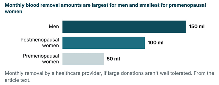
Figure 5. Monthly blood removal amounts by group, for people who don’t tolerate large donations well.
Most people can do this safely, but check your ferritin first, since removing blood isn’t appropriate if your iron levels are already low. Talk to your healthcare provider about whether this testing is appropriate for you.
Frequently Asked Questions (FAQs) About Microclots in Long COVID Blood
Q: What biological clues have researchers found behind long COVID symptoms?
A: Research shows that long COVID is strongly linked to fibrin microclots mixed with immune cell debris, especially neutrophil extracellular traps (NETs). These abnormal clots may restrict microcirculation and reduce oxygen delivery, which researchers think could contribute to fatigue, breathlessness, brain fog, and other persistent symptoms.
Q: How do NETs make long COVID microclots harder for the body to clear?
A: NETs form sticky webs of DNA and enzymes that bind to fibrin, forming clots that may resist normal breakdown. Because the body may struggle to dissolve them, these microclots may accumulate, prolonging vascular stress and maintaining symptoms for months or years.
Q: Does COVID increase clotting risk even after mild illness or recovery?
A: Yes. COVID can trigger inflammation, endothelial injury, and hypercoagulation. Clotting complications are most common in severe cases, but one large study found clot risk was still raised nearly a year after infection, including in people who were never hospitalized. Some people continue developing clots weeks to months after infection, which may contribute to post-recovery chest tightness, fatigue, and shortness of breath.
Q: Is long COVID rooted in mitochondrial dysfunction?
A: Long COVID may disrupt mitochondria through microvascular blockages, inflammation, and oxidative stress. This energy deficit may help explain fatigue, post-exertional crashes, and slow recovery, and is why many approaches focus on restoring mitochondrial function.
Q: What strategies can help support recovery from long COVID?
A: Potential approaches include proteolytic enzymes (which may help clear residual proteins), the I-RECOVER protocol, reducing linoleic acid intake to support mitochondrial health, using pharmaceutical-grade methylene blue under a healthcare provider’s supervision, and balancing iron and copper levels through diet or blood donation.
This article is for informational purposes only and does not constitute medical advice. Consult a qualified healthcare provider before making changes to your health regimen.